We can see objects as small as 0.1 millimeters, and that means we can just about see these lice eggs in our hair and tiny single-celled organisms like amoeba. But it's possible to see things much smaller than that if we use magnification.
There's three types of microscope: light, like this one here, and two types of electron microscopes, like those ones over there.
Light microscopes use light and mirrors and can see things as small as 400 nanometers. This allows us to get down to the world of the cell, and that means some pretty amazing things can be seen.
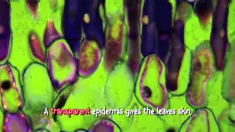
Here's an amoeba engulfing red blood cells and red and white blood cells moving through a tiny blood vessel. And human sperm. A leaf surface at 600 times magnification and the head of a dog tapeworm no bigger than a grain of rice. Plant cells crammed with chloroplasts. Look at these glucose crystals.
But light microscopes have a limit. Any object that's smaller than the wavelength of light appears blurred. But in the 1930s, a new kind of microscope was invented, which took our eyes further than they'd ever been before, to places we'd never seen before - the electron microscope.
The specimen is put in a vacuum and is viewed not by light waves but by a single beam of electrons that scans the surface, building up an image on a screen rather like a television picture. Because electrons have a wavelength 100,000 times smaller than light, electron microscopes can magnify objects up to 10 million times.
There are two types of electron microscopes: the transmission and the scanning. The scanning electron microscope scatters electrons across the surface of a specimen. It can magnify in incredible detail. This is a leaf surface under a scanning electron microscope. Both types of electron microscopes make black-and-white images, but these have been colourised to make them clearer and a lot more appealing to the eye.
Check out this fruit fly. A pubic louse and its claws. Cancer cells splitting. A blood clot. And human sperm cells on the surface of an egg.
But what about a transmission electron microscope? The difference with a transmission electron microscope is that it sees through things. It does this by sending beams of electrons, rather than light, through ultra-thin specimens. Using these microscopes, we're able to study the interior of cells and their organelles, and we've been able to get a better understanding of how pathogens, such as viruses, invade cells, like these HIV particles budding on the surface of a T cell.
Now a new type of electron microscope, a tunnelling electron microscope, has even made it possible to see the arrangement of atoms. Just how far will microscopy go?
Video summary
Science presenter Jon Chase describes three different types of microscope.
He explains how a light microscope works, and takes a look at sperm, red and white blood cells, and the surface of a leaf. He then discusses the difference between a transmission and scanning electron microscope and views some images of cells, organelles and virus particles under higher magnification.
Teacher Notes
Your class could use microscopes in a follow up activity to observe and draw the structure of magnified objects.
Interesting objects for students to look at could include different fabrics and the torn edge of paper. Pupils could also look at blood cells (providing an appropriate risk assessment has been completed).
These short films will be relevant for teaching biology and science in general at KS3 and KS4 in England, Wales and Northern Ireland and National 4/5 in Scotland.
Aerobic respiration. video
Science presenter Jon Chase explains aerobic respiration.
An explanation of photosynthesis. video
Jon Chase explains photosynthesis.
Enzymes and active sites. video
Jon Chase demonstrates the action of the enzyme catalase.
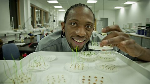
Factors that affect germination. video
Jon Chase investigates the effect of temperature, water and oxygen on seed germination.
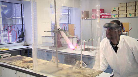
Food as fuel. video
A screaming jelly baby is demonstrated to show the energy content of food.
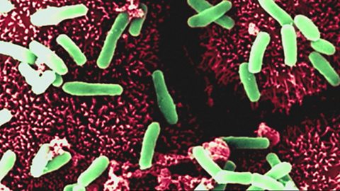
Microorganisms and bacteria. video
Personal possessions are swabbed for bacteria which are then cultured on agar plates.
Mitosis Rap. video
Jon Chase raps about the stages of mitosis.

Osmosis Rap. video
Science presenter Jon Chase raps about osmosis.
Photosynthesis Rap. video
Science presenter Jon Chase raps about photosynthesis.