Electron microscopes
How have light microscopes developed?
No-one knows who first invented the microscope but there have been key stages in their development:
- 1590s - Dutch spectacle makers Janssen experimented with putting lenses in tubes. They made the first A compound microscope uses two lenses, the objective lens and the eyepiece. The very short focal length objective lens produces a greatly-magnified image, then the short focal length eyepiece magnifies this further. . None of their microscopes have survived, but they are thought to have magnified from ×3 to ×9.
- 1660s - British scientist, Robert Hooke – also famous for his law of elasticity in Physics – observed and drew cells using a compound microscope.
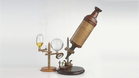
Image caption, A replica of Robert Hooke's compound microscope
1 of 2
- Late 1600s – Dutch scientist Antonie van Leeuwenhoek constructed a microscope with a single spherical lens. It magnified up to ×275.
- 1800s - the optical quality of lenses increased and microscopes are similar to the ones used today.
Throughout their development the magnification of light microscopes has increased, but very high magnifications are not possible. The maximum magnification with a light microscope is around ×1500 to ×2000.
The limits of the light microscope
The magnification of a microscope is not the only factor that's important when viewing cells. The detail that can be seen is also important.
The ability to see greater detail in an image depends on the The fineness of detail that can be seen in an image - the higher the resolution of an image, the more detail it holds. In computing terms, resolution is measured in dots per inch (dpi). or resolving power. This is the ability to see two points as two points, rather than merged into one.
Think about a digital photo. It can be enlarged but over a certain size, you won’t be able to see any more detail. It will just become blurry.
The resolution of a light microscope is around 0.2 μm, or 200 nm. This means that it cannot distinguish two points closer than 200 nm. One nm, or nanometre, is one billionth of a metre. This is written as:
\(\frac {1}{1~000~000~000}\) m, or in standard form as 1 × 10⁻⁹ m.
The electron microscope
Electron microscopes use a beam of electrons instead of light rays.
There are two types of electron microscope:
- The scanning electron microscope (SEM) has a large The distance between the nearest and farthest objects in focus. so it can be used to examine the surface structure of specimens. SEMs are often used at lower magnifications.
- The transmission electron microscope (TEM) is used to examine thin slices or sections of cells or A group of similar cells that carry out the same function, eg muscle tissue..
TEMs have a maximum magnification of around ×1,000,000, but images can be enlarged beyond that photographically. The limit of resolution of the transmission electron microscope is now less than 1 nm.
The TEM has revealed structures in cells that are not visible with the light microscope. We can now study cells in much finer detail.
