Looking at blood cells
You may be given some prepared slides of blood to examine with the microscope.
Many types of blood cell are 10 μm in size or less. You will need high power to examine them.
The slides will have been stained to show the cells, and cell features. The micrograph shows many red blood cells and three white blood cells.
An example of a commonly-used stain is Giemsa stain. It aids identification by staining:
| Red blood cells | Pink |
| Platelets | Pale pink |
| White blood cell cytoplasm | Pale blue |
| White blood cell nuclei | Magenta |
| Red blood cells |
| Pink |
| Platelets |
| Pale pink |
| White blood cell cytoplasm |
| Pale blue |
| White blood cell nuclei |
| Magenta |
Looking at cells in a blood smear
Aim
To use a light microscope to see blood cells in a smear.
Method
- Place a drop of blood onto a microscope slide.
- Add a drop of stain to the blood to make the cells easier to see.
- Carefully place a coverslip over the drop of blood. Sliding it slightly along the microscope slide will spread out the blood cells making them easier to see.
- Follow the instructions below to see the blood cells on a light microscope.
Rotate the In a compound microscope, the lens closest to the specimen has a very short focal length and produces a greatly magnified image of the specimen. so that the low power, eg ×10, is in line with the stage.
Turn the coarse focus so that the stage is as close to the objective lens as possible.
You should not look through the microscope to do this.
Place the microscope slide on the stage.
Line it up so that the specimen – if you can see it – is in the centre of the stage, where the light passes through.
Focus the slide towards you by turning the coarse focus adjustment.
Health and safety
Results
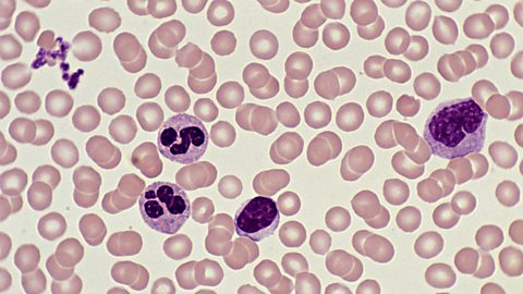
You are likely to see an image like this. Here you can see larger white blood cells and smaller red blood cells. The cells have been stained using Wright's solution stain.
