Microscopes
How have light microscopes developed?
No-one knows who first invented the microscope, but there have been key stages in their development:
- 1590s - Dutch spectacle makers Janssen experimented with putting lenses in tubes. They made the first A compound microscope uses two lenses, the objective lens and the eyepiece. The very short focal length objective lens produces a greatly-magnified image, then the short focal length eyepiece magnifies this further. . None of their microscopes have survived, but they are thought to have magnified from ×3 to ×9.
- 1650 - British scientist, Robert Hooke 1650 – also famous for his law of elasticity in Physics – observed and drew cells using a compound microscope.
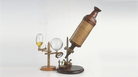
Image caption, A replica of Robert Hooke's compound microscope
1 of 2
End of image gallery
- Late 1600s – Dutch scientist Antonie van Leeuwenhoek constructed a microscope with a single spherical lens. It magnified up to ×275.
- 1800s - the optical quality of lenses increased and the microscopes are similar to the ones we use today.
Throughout their development, the magnification of light microscopes has increased, but very high magnifications are not possible. The maximum magnification with a light microscope is around ×1500.
